Muscle Structure Heterogeneity
Overview
Muscle structures have been shown to vary spatially within the muscle. This spatial heterogeneity has been explored in the diaphragm where blood flow, cross sectional area, connective tissue amount, and neuromuscular junction spacing vary regionally. As a side project, I developed a whole mount technique for imaging diaphragm regions which proved useful in studying structural heterogeneity. Our first study explored sarcomere variability across diaphragm regions. Sarcomere length is a fundamental parameter of skeletal muscles. Additionally, muscle damage or diseases can cause a disruption in sarcomere organization and impair muscle function.

Diaphragm sarcomere organization
My largest study on sarcomere organization has been done in collaboration with Bridget Ward and Catherine Henry, undergraduate students at the University of Virginia. The diaphragm is a unique circular muscle, with radial muscle fibers spanning from the chest wall to the central tendon. We hypothesized that the regions of the diaphragm (ventral, mid costal, and dorsal) had varying sarcomere lengths. We found the sarcomere lengths of the diaphragm regions were different in healthy mice. Additionally, there were sarcomere differences between healthy and mdx mice, a model of Duchenne muscular dystrophy.

Catherine has taken this study even further by including 1) multiple age groups and 2) expanding the muscle parameters measured to investigate fiber branching, amount of interstitial space, muscle fiber cross-sectional area, and macrophage counts. Together, we have found regions of the diaphragm differ with respect to common mdx disease progression markers, like centrally located nuclei or macrophage counts. This work was published in PloS one (see my publications page for a link).
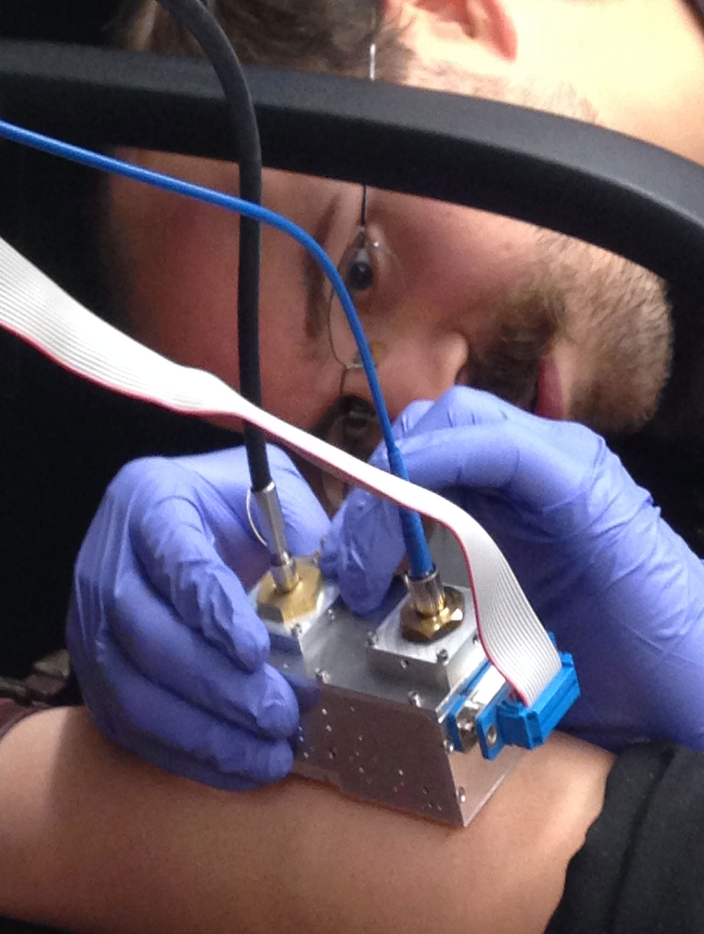
In vivo sarcomere assessment
